Introduction
Medical imaging has revolutionized the diagnosis and management of kidney-related conditions. Among the various techniques, computed tomography (CT) has emerged as a key tool in assessing abnormalities in renal anatomy and function. One of the conditions often evaluated using this technology is cortical thinning kidney CT, which refers to the use of CT scans to assess the thinning of the renal cortex. This article explores the significance, causes, diagnostic process, and implications of cortical thinning in kidney CT scans, ensuring a comprehensive understanding of this important topic.
What is Cortical Thinning in the Kidney?
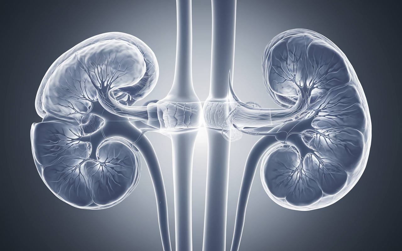
Cortical thinning refers to the reduction in the thickness of the kidney’s outer layer, known as the renal cortex. The cortex plays a vital role in the kidney’s filtration process, as it contains the glomeruli and a significant portion of the nephron. When the cortex becomes abnormally thin, it can indicate various underlying conditions or chronic damage to the kidney tissue.
Cortical thinning kidney CT is a diagnostic approach used to visualize and measure the extent of cortical thinning. Through this imaging technique, healthcare providers can detect subtle structural changes and identify potential causes of kidney dysfunction.

Causes of Cortical Thinning in Kidneys
The causes of cortical thinning are diverse and often linked to chronic or progressive kidney conditions. Common contributing factors include:
- Chronic Kidney Disease (CKD): One of the most frequent causes of cortical thinning observed in cortical thinning kidney CT is CKD. Prolonged damage to the kidneys leads to scarring and atrophy, manifesting as a thinning cortex.
- Hypertension: Chronic high blood pressure can damage the small blood vessels in the kidney, reducing blood flow and causing cortical thinning.
- Diabetes Mellitus: Diabetes is a leading cause of kidney damage. Hyperglycemia and related complications can contribute to structural abnormalities, including cortical thinning, as observed on cortical thinning kidney CT scans.
- Obstruction or Reflux Nephropathy: Conditions such as urinary tract obstruction or vesicoureteral reflux can lead to cortical scarring and thinning over time.
- Infections: Recurrent kidney infections (pyelonephritis) may result in localized cortical thinning due to inflammation and tissue destruction.
Role of CT Scans in Diagnosing Cortical Thinning
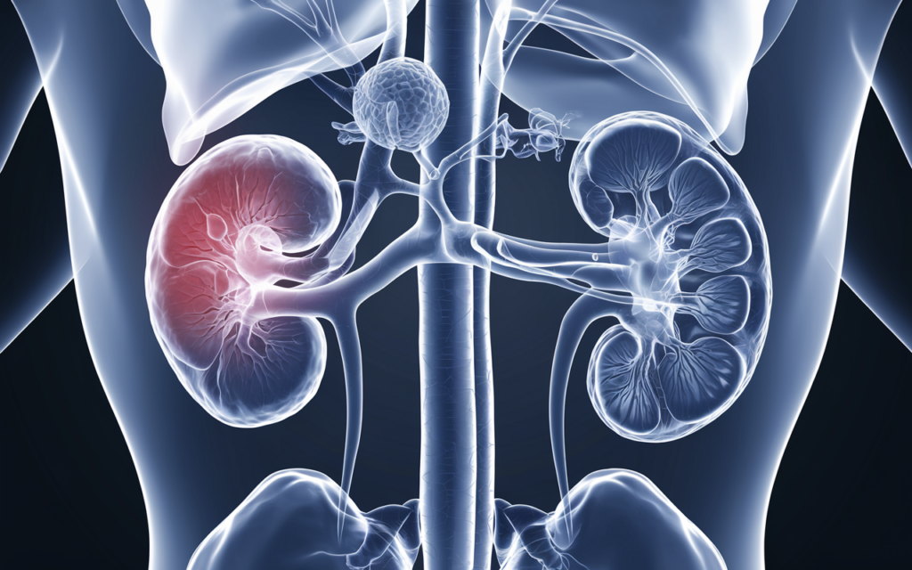
The use of cortical thinning kidney CT provides a detailed and accurate visualization of the kidneys, allowing for precise measurement of cortical thickness. CT scans use X-ray technology to generate cross-sectional images, which can be reconstructed into 3D models for enhanced evaluation.
Advantages of Using CT Scans for Cortical Thinning
- High Resolution: CT scans provide high-resolution images, enabling the detection of subtle changes in cortical thickness.
- Quantitative Measurement: The thickness of the renal cortex can be quantitatively measured, offering objective data to monitor disease progression.
- Identification of Associated Conditions: A CT scan can reveal other abnormalities, such as kidney stones, masses, or cysts, that may contribute to cortical thinning.
- Non-invasive and Quick: While CT imaging involves radiation, it remains a non-invasive and relatively fast procedure for evaluating renal conditions.
In cortical thinning kidney CT, specific protocols may be employed to enhance visualization. Contrast-enhanced CT scans, for instance, can improve the differentiation between the renal cortex, medulla, and surrounding structures.
Assessment Techniques in Cortical Thinning Kidney CT
Several parameters are analyzed during a cortical-thinning kidney CT scan. These include:
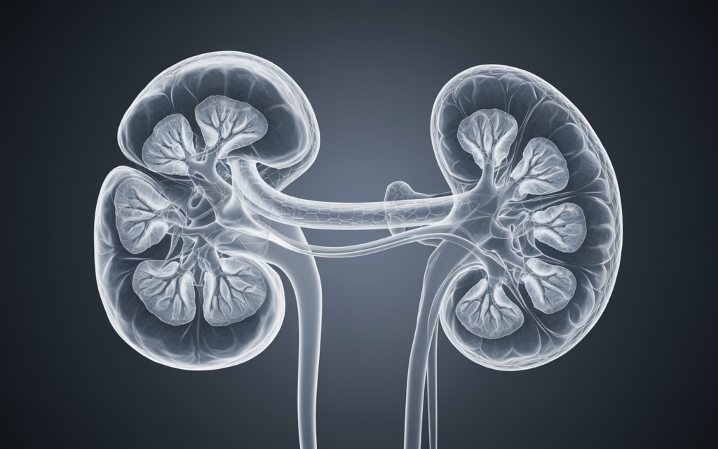
- Cortical Thickness Measurement: The thickness of the cortex is measured at various points, typically at the poles and the mid-zone of the kidney. Values below the normal range suggest thinning.
- Corticomedullary Differentiation: In healthy kidneys, there is a clear distinction between the cortex and medulla. Loss of this differentiation may accompany cortical thinning.
- Kidney Size and Shape: CT scans can detect changes in kidney size and contour, which may correlate with cortical thinning and underlying pathologies.
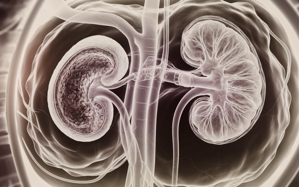
- Scarring or Atrophy: Evidence of scarring or focal atrophy is often seen alongside cortical thinning and can provide clues about the underlying cause.
Clinical Implications of Cortical Thinning
Cortical thinning observed in cortical thinning kidney CT has significant clinical implications. It often indicates reduced functional nephron mass, which can impact the kidney’s ability to filter blood effectively. Depending on the extent of cortical thinning and the underlying cause, the prognosis may vary.
Associated Risks
- Progression to End-Stage Renal Disease (ESRD): Advanced cortical thinning is often associated with irreversible damage, potentially leading to ESRD.
- Impaired Kidney Function: Patients with cortical thinning may experience reduced glomerular filtration rate (GFR), reflecting impaired kidney function.
- Increased Risk of Complications: Chronic kidney conditions, including cortical thinning, can increase the risk of cardiovascular disease and metabolic disturbances.
Monitoring and Management
Regular imaging and functional assessments are crucial for patients with cortical thinning. In addition to cortical thinning, kidney CT, blood tests, urine analysis, and other imaging modalities may be employed to monitor disease progression and guide treatment decisions.
Treatment and Management Strategies
The management of cortical thinning depends on the underlying cause and the extent of kidney damage. While the condition itself cannot be reversed, interventions can slow its progression and mitigate complications.
- Controlling Blood Pressure: Hypertension is a major contributor to kidney damage. Medications such as ACE inhibitors or ARBs are commonly prescribed to manage blood pressure and protect kidney function.
- Managing Diabetes: Tight glycemic control can prevent further damage in diabetic patients with cortical thinning, as indicated on cortical thinning kidney CT scans.
- Addressing Obstructions: Surgical or medical interventions may be necessary to resolve obstructions or reflux contributing to cortical thinning.
- Lifestyle Modifications: Dietary changes, exercise, and smoking cessation can improve overall kidney health and reduce the burden of contributing risk factors.
- Renal Replacement Therapy: In cases where cortical thinning progresses to ESRD, dialysis or kidney transplantation may be required.
Prognosis and Long-Term Outlook
The prognosis for patients with cortical thinning depends on several factors, including the underlying cause, the extent of thinning observed on cortical thinning kidney CT, and the patient’s overall health. Early detection and proactive management can significantly improve outcomes, emphasizing the importance of routine monitoring in at-risk populations.
Future Perspectives on Cortical Thinning Kidney CT
Advancements in imaging technology are poised to enhance the evaluation of cortical thinning. Techniques such as functional CT imaging, artificial intelligence (AI)-driven analysis, and integration with other modalities like magnetic resonance imaging (MRI) may provide deeper insights into the pathophysiology of cortical thinning. These innovations could also lead to earlier detection and more personalized treatment strategies.
Moreover, research into regenerative medicine and nephron repair holds promise for addressing cortical thinning at its core. Until then, cortical thinning kidney CT remains an indispensable tool in diagnosing and managing this condition.
Conclusion
Cortical thinning kidney CT is a vital diagnostic approach that enables healthcare providers to assess and monitor the thinning of the renal cortex effectively. By identifying the underlying causes, such as CKD, hypertension, diabetes, and infections, clinicians can implement targeted interventions to slow disease progression and improve patient outcomes.
This imaging technique not only provides detailed anatomical insights but also guides treatment decisions and helps predict the prognosis. As technology continues to evolve, the role of cortical thinning kidney CT will undoubtedly expand, offering new possibilities for understanding and managing kidney conditions. Through early detection and comprehensive care, patients with cortical thinning can achieve a better quality of life and reduced risk of complications.